
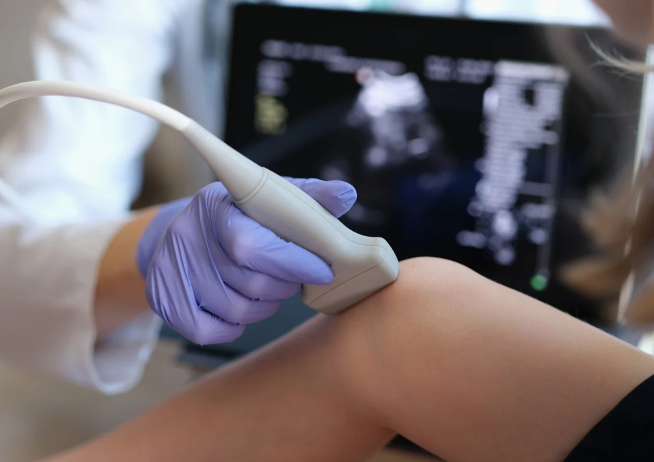
How to choose an ultrasound device: a practical guide

Come scegliere un ecografo in fisioterapia: guida pratica

Musculoskeletal ultrasound is now an essential tool in physiotherapy, for both functional assessment and treatment monitoring. But with so much choice on the market, how do you choose an ultrasound device that suits your clinical needs and that you can rely on? 

Price and appearance are not enough: technical specifications, advanced features and usability all directly affect the quality of care. Here are the key factors to consider.
  • Image resolution and depth
  • Imaging modes and advanced features
  • Ergonomics, portability and reliability 
  • Want to know more or receive commercial offers?

Image resolution and depth

Image quality is the deciding factor, as it determines how accurately anatomical structures can be identified and changes detected. Two parameters are critical.

Resolution is the level of detail in the image. High resolution makes it possible to clearly distinguish muscle fibres, tendons, ligaments and nerve fascicles, and to pick up subtle changes in echogenicity or fibrillar continuity that shape clinical interpretation and treatment planning.

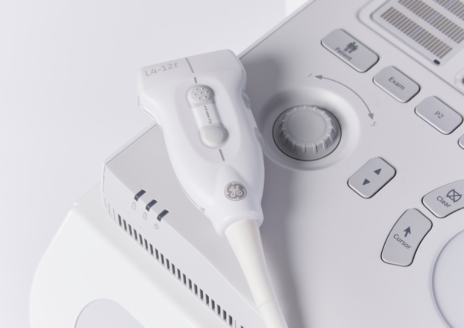
Depth depends on transducer frequency. High-frequency probes (10–18 MHz) provide excellent resolution for superficial structures, while lower-frequency ones (5–8 MHz) reach deeper planes at the cost of some sharpness. As different body regions call for different settings, look for a machine that allows frequency and depth to be adjusted flexibly: this is what makes scans reliable and reproducible over time.

Imaging modes and advanced features

B-mode, the most widely used mode, produces real-time two-dimensional images of muscles, tendons and ligaments. M-mode is useful for analysing dynamic movement, such as tendon gliding or diaphragmatic excursion.

Doppler assesses tissue vascularity, which is valuable in inflammatory conditions, tendinopathies and the monitoring of tissue repair. Colour and power Doppler improve sensitivity to low-velocity flow, which helps in chronic or microvascular lesions.

Elastography measures tissue stiffness. It is useful for assessing fibrosis and scarring and for monitoring treatments that target soft-tissue elasticity. Not every machine has it, but it makes assessment more objective and quantifiable.

Come scegliere un ecografo in fisioterapia: guida pratica

Ergonomics, portability and reliability

A compact, intuitive device with well-placed controls and quick adjustments makes scanning more efficient and reduces practitioner fatigue. Light weight and good battery life matter most for home visits, small treatment rooms and sports settings. Finally, consider the quality of technical support, software updates and the long-term availability of compatible probes. Choosing an ultrasound device for physiotherapy means striking the right balance between image quality, advanced features and practical considerations, so that it meets today’s needs and grows with your practice. 

Want to know more or receive commercial offers?

To know more, write at info@easytechitalia.com or call 055 8455216.

Previous Post
Anterior cruciate ligament injury: how to achieve the best recovery
Other articles